Colic in horses: Even though colic is a common ailment in horses, it can be mysterious and frightening for horse owners. Colic is a generalised term referring to any type of abdominal pain in the horse and most commonly originates from within the gastrointestinal tract, or gut. This article will focus on the gastrointestinal causes of colic to help horse owners broaden their understanding of the problem and be able to make informed decisions regarding their horse if colic does occur.
Signs of Colic in Horses
The clinical signs of colic can vary greatly from horse to horse. The most common symptoms include:
- Lying down,
- Rolling on the ground,
- Kicking at the abdomen,
- Flank watching (looking at abdomen),
- Pawing at the ground,
- Sitting like a dog,
- A swollen belly,
- Stretching out with the hind legs,
- Lack of appetite,
- A decrease in manure output, and
- Development of diarrhoea.
Horses may exhibit one or several of these clinical signs at the same time.
Watch this video to see some of the classic signs:
Mild colic signs can quickly progress and become more severe in a short period of time. However, some horses are stoic; therefore, the severity of the clinical signs does not always coincide with the severity of the problem.
Call your vet
Early intervention and treatment of a horse with colic will increase the chance of having a favourable outcome. It is important to seek veterinary advice if your horse is showing any of the above clinical signs. A veterinarian can advise on the situation to determine if your horse may be monitored first or will require immediate veterinary attention.
Your vet may attend your horse on the farm or your horse may be required to be transported to a veterinary hospital for examination. For farm visits, it is important to monitor your horse until your vet arrives.
This includes not just monitoring their clinical signs, but also placing your horse in an area free of obstacles, including fences, equipment, or small and confined spaces, and/or by walking them to prevent injury. If your horse is lying down and rolling, it is essential to monitor them and get them up safely.
Food should be withheld until your vet arrives. If loading your horse for travel to a veterinary clinic, your vet can give you advice on any treatment prior to loading.
When your vet arrives
To start with, they will take a thorough history by asking a series of questions, which may include:
- What symptoms is the horse showing?
- How long has the horse been showing these signs?
- How frequently are they passing manure? What is the consistency of the manure – soft and mushy or hard and dry?
- How much water are they drinking?
- When was the horse last wormed?
- If they were wormed recently, what wormer was used?
- What is their dental history?
- Has there been any change in feed, pasture or housing?
- Has the horse previously had colic?
- Has the horse had any treatment or medications?
- Is the horse insured for veterinary treatment?
- Are there any other horses on the property showing similar clinical signs?
- Is there any other relevant history pertinent to the horse?
Following taking a history, your vet will perform a physical examination of your horse. This includes checking the eyes and colour of the gums, listening to the heart, lungs and gut sounds with a stethoscope, checking the legs and feet, as well as taking a temperature. Further diagnostic tests may be recommended based on their initial findings.
Once the external areas of the body are checked, your vet may pass a tube through the nose of your horse and into the stomach (nasogastric tube).
If the stomach is enlarged from fluid or gas buildup, the tube allows the fluid to escape and gives the horse some temporary relief.
Fluid or gas buildup may occur in the stomach due to partial or complete obstruction of the intestines further down the line. Your vet may then be able to directly medicate your horse through the tube if treatment (drench) is required.
Diagnostic tests
Drawing a sample of blood and testing it for different factors can help determine if your horse has any signs of infection (haematology), inflammation (fibrinogen and serum amyloid A), organ problems (biochemistry), dehydration or shock (haematocrit and total protein), and abnormal electrolyte values (not eating or lost via diarrhoea).
An abdominal ultrasound can be very helpful in determining the state of the intestines. If the intestines are distended with gas (twisted or blocked), thickened (don’t absorb nutrients very well), not moving (decreased motility/ileus) or have abnormal structures which could be causing a problem (abscesses or tumours), then these may be detected with an ultrasound examination.
An increase in the amount of fluid in the abdomen can also be seen and may indicate infection or inflammation within the abdomen (peritonitis), or abnormal positioning of the intestines (twists, rupture or entrapments). If there is increased fluid in the abdomen, your vet will likely suggest taking a sample of that fluid (abdominocentesis) to check its colour, amount and if there is infection present (fluid analysis).
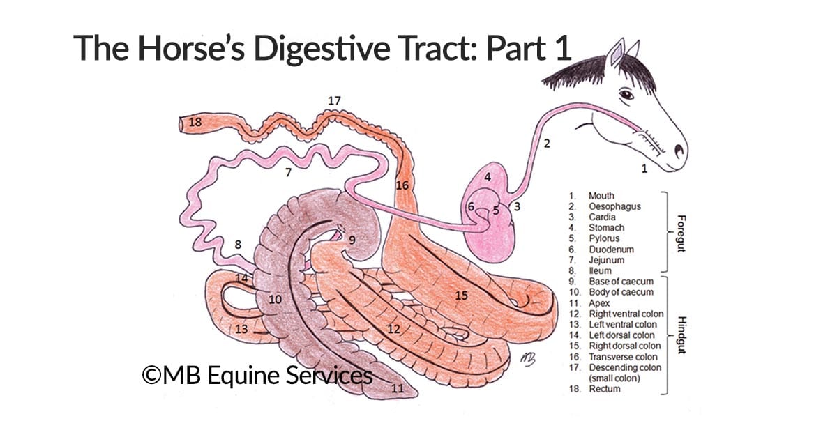
A rectal examination might be the next diagnostic step to investigate colic symptoms. Your vet will carefully palpate the abdomen from the inside by inserting their hand and arm into the rectum of your horse. This will help them to determine if the intestines, colon or caecum are in the wrong place (displacement), and check for gas expansion of the intestines. They may also collect a sample of faeces at the same time to perform a faecal egg count to determine if there is any worm burden.
Colic causes
There are numerous causes and types of colic that can be seen in horses:
- Partial or complete obstruction of the large or small intestine, which includes feed/sand impaction, foreign bodies, faecal stones (enteroliths), intussusception or parasites.
- Spasmodic colic, which is irregular (spasmodic) contractility of the gastrointestinal tract.
- Strangulating obstruction (the gut is twisted around cutting off the blood supply to the intestine), which includes small or large intestinal torsion/volvulus, or a pedunculated lipoma (fatty tumour on a long piece of tissue, which wraps around the gut like a piece of string cutting off the blood supply).
- Displacements where the intestines have moved to the wrong anatomical location. Dorsal displacements of the colon or nephrosplenic entrapments (gut trapped between tissue stretching from the spleen to the kidney).
- Inflammation of the small intestine (enteritis), large intestine (colitis) or stomach ulcers.
- A tumour or abscess attached to the intestinal wall or encompassing the intestines.
A horse may have one type of colic or many at the same time. Despite numerous diagnostic tests available, there are colic cases where a definite cause is never found.
Treatment of colic can be medical or surgical, depending on the type and severity of the clinical signs.
Most suspect impactions, enteritis, colitis and, occasionally, displacements can be resolved with medical treatment. This may be basic therapy (pain relief and drenching) or complex (pain relief, fluid therapy in the vein, antibiotics, plasma therapy and hospitalisation).
Pain relief may be in the form of anti-inflammatories, sedatives and/or opioids. A drench may be administered through a nasogastric tube as previously detailed and can include a combination of electrolytes, a faecal softener or paraffin oil, and water.
The aim of a drench is to correct any dehydration (encourage drinking) and soften the intestinal contents to encourage easier passage. Initial basic medical treatment may be performed on the farm.
In severe cases of colic, more complex therapy may be required, often at a veterinary clinic. Hospitalisation allows for constant monitoring and continuous fluid therapy. Continuous fluid therapy rehydrates your horse, and can soften gut contents or maintain fluid in the body if there is diarrhoea.
Medication may be given to support gastrointestinal motility, provide additional or constant pain relief, treat infections (antibiotics), prevent ulcers, and replace any electrolytes lost from not eating or due to diarrhoea.
Surgical treatment of colic is necessary in cases where there is severe pain or the horse fails to respond to medical treatment.
Colic surgery
Surgery (exploratory laparotomy) is necessary to correct severe impactions, displacements, torsion/volvulus, or remove dead bowel or tumours.
Post-operative care is aimed at supporting the healing process with medical treatment and slow re-introduction of feed. In some instances, there may be complications to recovery from surgery.
These may include decreased intestinal motility (ileus), infections of the surgery site, the intestines adhering together (adhesions) or infection inside the abdomen (peritonitis).
Horses without post-operative complications are generally able to go home within 5-7 days following surgery. The survival rate for horses undergoing colic surgery and recovering from anesthesia can be as high as between 80 and 90%, depending upon the cause of the colic, and most go on to lead successful careers as show horses, racehorses or beloved riding horses.
Talk with your vet
Recognising the clinical signs of colic and receiving prompt veterinary attention can be the key to a horse recovering quickly with the least amount of pain. Having a good working relationship with an equine veterinarian is helpful in choosing the level of care, diagnostic tests and treatment for any horse with colic, and will increase the chance of having a favorable outcome to any episode.