Laminitis… A condition that strikes fear into every horse owner.
Long recognised as a cause of lameness, laminitis can have debilitating long-term effects on horses and ponies. It often becomes an insidious, chronic issue that requires careful management, including a constant evaluation of the diet and specialised hoof care.
Despite its widespread prevalence, especially amongst native and pony breeds, and despite a very large and impressive body of scientific study, we still don’t have a clear picture of all the modalities, triggers nor the progression of this painful condition.
Laminitis is still an enigma and a fascinating area of research that offers scientists opportunities for making important breakthroughs in understanding that may soon lead to new treatments.
Earlier this year, The Veterinary Journal published a comprehensive review of ‘what we know so far’ and how our understanding of laminitis is changing as science continues to provide a better and more complete picture.
‘Paradigm shifts in understanding equine laminitis’ by Janet Patterson-Kane, Ninja Karikoski and Professor Cathy McGowan is a thorough literature review and shows where the future of laminitis research should be heading.
Historically, ‘classic’ laminitis was linked to systemic inflammatory response syndrome (SIRS) most often related to carbohydrate overload of the gastrointestinal tract, however, it is now recognised that almost 90% of laminitis cases are caused by endocrine (hormonal) disease – high insulin levels.
An example of SIRS caused laminitis occurs when the horse gets into the feed room or breaks into a lush, spring grass paddock and gorges on carbohydrate rich feed and plants.
When this happens, the small intestine is overwhelmed and loses its capacity to digest the carbohydrate component which flows into the large intestine where it is rapidly fermented by the bacteria, fungi and protozoa that inhabit the space. The acidic environment that results (acidosis) triggers an inflammatory chain of events that has been studied extensively and results in the well-known clinical signs of laminitis.

Anatomy of the equine hoof showing the laminae in detail. Image ©MB Equine Services.
Other causes of SIRS have also been described (but studied less thoroughly), including metritis (inflammation of the uterine wall in broodmares) as well as supporting limb lameness.
Today we know the vast majority of laminitis cases are linked with hyperinsulenaemia (high insulin). This was first hypothesised in the 1980s when researchers noticed that insulin resistance was commonly associated with laminitis, especially in pony breeds, and led to the description of ‘endocrinopathic laminitis’.
Despite this discovery, researchers continued investigating the SIRS model of laminitis until 2006, when a ground breaking study found a fivefold increase in baseline blood insulin levels at the onset of a carbohydrate induced laminitis episode (Treiber et al., 2006).
Meanwhile, Prof. Cathy McGowan’s team published another important paper emphasising hormonal-related laminitis after studying horses with pituitary pars intermedia dysfunction (PPID, formerly known as ‘Cushings’) and other endocrine (hormonal) diseases now known as equine metabolic syndrome (EMS).
By studying the clinical findings, they were able to develop an experimental model of laminitis resulting from consistently elevated blood levels of insulin (hyperinsulinaemic laminitis).
More recent studies have suggested that the clinical management of laminitis, particularly as a result of the new knowledge of spontaneous endocrinopathic laminitis (laminitis that is apparently brought-on by nothing other than abnormal insulin regulation) warrants re-evaluation.
So, where does this lead?
A research review published in The Veterinary Journal outlines three major shifts in thinking about laminitis which are important for veterinarians and clinicians to understand in order to offer the most suitable treatment plan.
1. Laminitis is a clinical syndrome (not a disease)
Laminitis is no longer described as a ‘discrete disease process’, but rather, a clinical syndrome, that is, a collection or set of signs and symptoms that characterise a disease process.
In the case of laminitis, the symptoms can result from several systemic diseases, from opposing limb lameness and from endocrine disease.
The change of description from disease process to syndrome might seem unimportant, but in veterinary and research terms it’s a game changer. It comes as a result of multiple studies repeating the results of Asplin et al. the research team overseen by Prof. Cathy McGowan who showed in 2007 that laminitis could be induced in 5 out of 5 previously healthy horses following exposure to prolonged periods of increased blood insulin levels.
This means that in order to implement suitable management and give realistic prognosis of prevention and recurrence of laminitis, veterinarians must first make an accurate diagnosis of the associated causes and disease.
To treat endocrinopathic laminitis we must understand how to control insulin levels and exactly how insulin causes hoof tissue damage.
2. Endocrinopathic laminitis predominates in lame horses
Hormone related laminitis is now understood to be the most common form of naturally occurring laminitis in developed countries.
Earlier, there was a misunderstanding that laminitis was predominantly the result of SIRS because of an apparent prevalence of SIRS type laminitis seen in lame horses presented at veterinary referral hospitals.
When further research conducted amongst the general population of horses and ponies showed that SIRS type laminitis represented only 12% of the laminitic cases reported by horse owners, the interest in the new model of laminitis caused by insulin disregulation and hyperinsulinemia grew.
Further studies corroborated these findings, identifying endocrinopathic laminitis in 90% of horses and ponies presenting for lameness. It became clear that the SIRS model of laminitis was less prevalent than originally thought and that future research should concentrate on hormone-related laminitis.
We now know that the major hormonal disorders associated with laminitis are EMS and PPID.
Field studies have shown that the prevalence of hyperinsulinaemia and laminitis is greater in horses with PPID than in age-matched controls. However EMS is emerging as the predominant endocrine cause of laminitis.
The next frontier will be controlling insulin levels in horses reliably, and early trials are being conducted with drugs currently used in humans.
At this stage, the best recommendations for lowering insulin are medication for PPID horses, as much exercise as possible and a diet that is lower than 10% in sugar and (ESC) starch.
3. The pathology of laminitis
Are endocrinopathic and SIRS associated laminitis different or is it just a matter of degree?
Lastly, the paper explains that our understanding of the pathology (causes and effects) of laminitis has changed with the identification of lamellar cell stretch as a potentially early warning sign.
Researchers have compared the effects on the hooves at a cellular level from both, SIRS and endocrinopathic laminitis affected horses.
The progression of disease is different in each type since SIRS associated laminitis is often rapid-onset and follows a distinct and often severe pattern of disease, while endocrinopathic laminitis is often slow-onset, becoming chronic having a long phase of development before clinical symptoms are seen.
Scientists studying the cells of hoof tissue in affected individuals found that the lesions associated with either type of laminitis were not particularly marked but were similar for each type.
They did, however, add a ‘disclaimer’ saying that the endocrinopathic laminitis experimental (artificially induced) model requires a sudden increase in blood insulin levels and this may differ from the gradual increase over time which occurs with naturally occurring endocrinopathic laminitis. Nevertheless, the lamellar structures appear to be an early target in the disease process.

IMAGES A & B show normal or ‘standard’ lamellae of the equine hoof under microscope. Note the orientation of the lamellae ‘fingers’ is more perpendicular. IMAGES C & D show the contrasting stretching and acute angling that has been associated with laminitic lamellae. (PEL stands for primary epidermal lamellae and SEL is the secondary epidermal lamellae.) Images sourced from The Veterinary Journal.
The study of the cellular structures of the hoof and the possible development of a ‘grading’ system to classify the severity of laminitis is made even more complicated by the fact that there appears to be a large degree of natural variation even in ‘healthy’ ‘normal’ feet. Without having defined a ‘normal’ with which to compare, it is very difficult to contrast cytological (cell) samples.
Early changes at cellular level that have been noted in both SIRS and endocrinopathic laminitis include a veering away from the perpendicular (up-down) orientation of the secondary epidermal lamellar nuclei relative to their basement membranes.
This means that the cells shift their orientation, with their nuclei (central core) becoming more centralised within the cell. The cells were noted to lengthen, narrow and develop tapered tips, making it difficult to distinguish the primary from the secondary epidermal lamellae.
This stretching of the cells is now thought to be a key early event in the onset of laminitis and might be the primary event leading to the later symptoms noted by the observer.
Changes ahead
The paper asks whether, with what we now know, the terminology itself should be changed to reflect the new understanding.
Whilst laminitis caused by carbohydrate overload shows some significant indicators of inflammation, these same pointers do not exist in hormone related laminitis. In fact, endocrinopathic laminitis is associated with minimal inflammation, unlike the SIRS model.
Furthermore, the amount of inflammation seen at a cellular level with either type of laminitis is far less than that seen in many other tissues undergoing similar stress and compromise.
The question then is whether the suffix ‘itis’ (meaning inflammation) is even warranted for the syndrome. The authors ask if it might be more appropriate for the term ‘laminopathy’ to be used for endocrinopathic laminitis.
Before the model for endocrinopathic laminitis had been fully developed, the research focus and primary damage was generally accepted to be a failure of the basement membrane (BM) within the hoof capsule.
This membrane (which effectively intersects and ‘joins’ the primary and secondary lamellae) was thought to be compromised leading to the partial or complete failure of the lamellar attachment and the ‘degloving’ of the hoof which most of us associate with the disease process of laminitis.
However, BM separation was not found in all of the carbohydrate overload-based models, being still intact and attached during the acute phase of the process in one study. It has also been noted that in most hyperinsulinaemic models, the BM damage was minimal.
The research has failed to show a solid primary role of BM pathology and, although there is no question that is plays a role in the cascade of lesions following initial onset, there is now strong debate about the role of BM breakdown as a primary contributor to either SIRS or endocrinopathic laminitis.
It is important to note, that the only available way to study the inner structures of hooves affected by SIRS and endocrinopathic laminitis thus far, has been in an experimental setting. In this setting, horses and ponies are artificially subjected to either a digestive system overload of carbs or a rapid-onset blood elevation of insulin. These can only somewhat imitate the effects which might be seen in naturally occurring endocrinopathic laminitis.
In 2015, the histopathology (cellular abnormalities) of 14 cases of naturally occurring endocrinopathic laminitis were compared with 25 age and breed matched controls (Karikoski et al.).
Microscopic areas of abnormal change (lesions) were noted close to the hoof wall. Cell death, lamellar fusion, cell proliferation and partial replacement with unusual keratinised material were all recorded in the samples of the laminitic horses. In some horses, there was evidence of tearing close to the hoof wall which invariably involved the BM, primary and secondary epidermal lamellae, compared to SIRS induced laminitis where tearing was seen to exclusively involve the BM and was at a deeper level of the lamellar structure.
Whilst these findings are interesting and not yet fully understood, it remains unclear whether naturally occurring endocrinopathic laminitis and SIRS-associated laminitis are fundamentally different diseases.
The good news
What is very encouraging about all this work is that, chronically developing hormonal laminitis, seems to have a protracted pre-clinical stage (a time where the disease process is in force but the clinical symptoms have not yet appeared). This affords us a good window of opportunity for intervention and preventative management.
Divergent hoof rings were seen in all but one of the 14 horses studied. By intervening at the early stage – when hoof rings are noted – the prevention of the critical lamellar stretching and associated pathology may be possible.

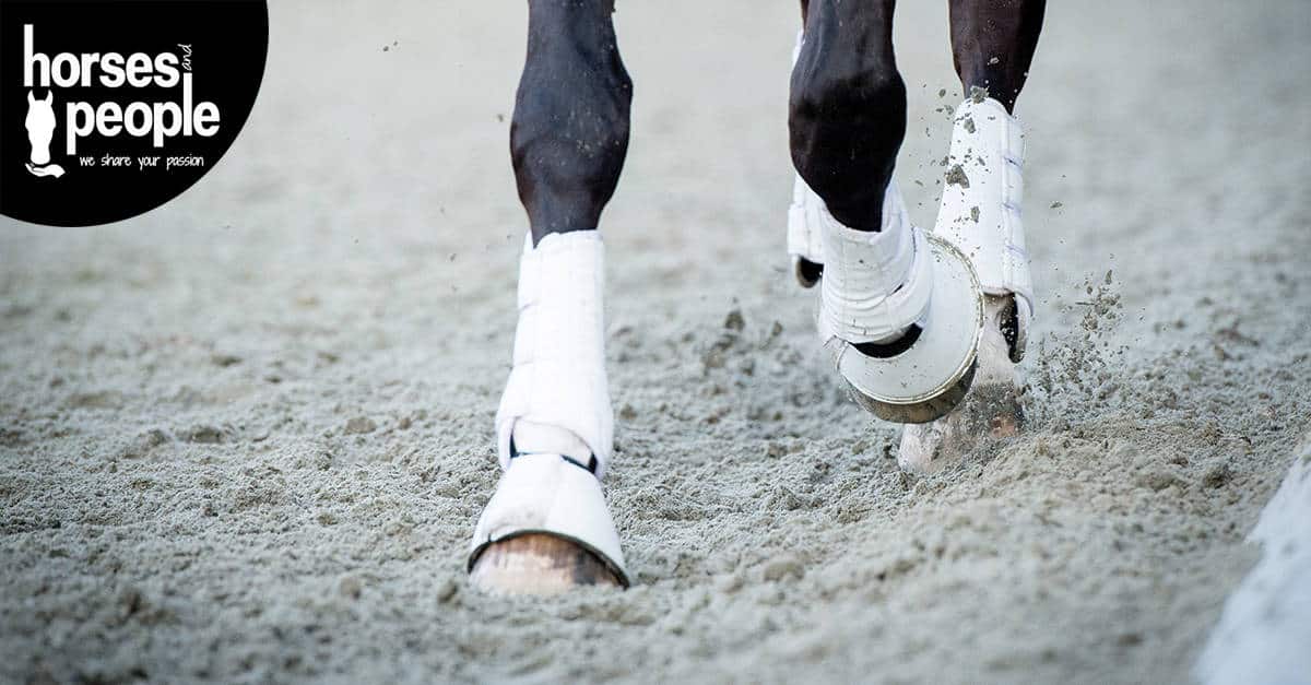
The tell-tale signs of a laminitic hoof – regardless whether the horse seems sound, don’t delay act now! Photo by Cristina Wilkins.
The future
Future research will focus on the mechanisms of laminitis at the cellular level, investigating the relationship between insulin and cellular changes, as well as possible intervention methods at critical time periods during the acute phase of laminitis.
Lamellar epithelial cell stretching as a key early event will be the focus of future studies investigating how and why this stretching occurs and whether it is preventable. The possibility of preventative drugs and treatment regimens is still beyond reach, but every study brings us that little bit closer to the day that laminitis becomes a manageable and preventable condition.
This paper is important because it highlights recent advances in our understanding of laminitis and suggests future research avenues to continue our understanding.
Laminitis is now considered to be a syndrome most commonly resulting from endocrine diseases such as PPID and EMS.
There is a long period of time where no symptoms of laminitis are evident but can be noticed by the development of divergent hoof rings.
Noticing these hoof rings gives the owner, hoof care professional and veterinarian a window of opportunity to intervene with treatments where appropriate, exercise, diet and management changes.
There is still a lot to learn though, so it is hoped that laminitis will continue to receive scientific attention.
Reference
The article titled: Paradigm shifts in understanding equine laminitis by J.C. Patterson-Kane, N.P. Karikoski and C.M. McGowan is open access and available on this link: https://doi.org/10.1016/j.tvjl.2017.11.011